Perceptive Imaging is a leading imaging core lab with the experience and ability to provide a wide range of services involving neuroimaging in clinical trials. Perceptive CNS group understands the unique and custom requirements of Multiple Sclerosis (MS) trials and has the flexibility, creativity, bandwidth, and understanding already in place to effectively manage and support these clinical trials.
A significant portion of our CNS experience is in Multiple Sclerosis (MS). In addition to standard MS assessments such as lesion counts (including lesion evolution), lesion and brain volumes, Perceptive also supports advanced MS analyses.
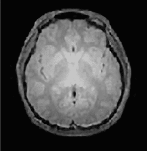
Magnetization Transfer Imaging (MTI)
Magnetization Transfer Imaging is a method that has been found to correlate with clinical disability in MS patients. This method is sensitive to the extent of myelination and provides a quantitative assessment of myelin integrity. The MT ratio, a parameter derived from MT imaging data, is a useful imaging biomarker for assessing extent of demyelination and remyelination in MS clinical studies.
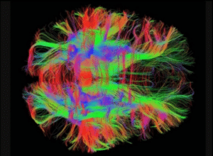
Diffusion Tensor Imaging (DTI)
DTI has been found to provide information about tissue microstructure and architecture including size, shape and organization and as a result represents an effective tool for evaluating white matter tissue integrity. The DTI tool can be used to obtain useful information in many ways – evaluation of Gadolinium-enhancing lesions by DTI provides information about tissue changes in active lesions; analysis of normal appearing white matter (NAWM) using the DTI tool may provide some insight into the early changes in the white matter (WM) in MS. Parameters such as mean diffusivity (MD), fractional anisotropy (FA) and volume ratio (VR) are some of the most important quantitative indices that are extracted from DTI data.
Functional Magnetic Resonance Imaging (FMRI)
fMRI can be used to measure brain activity at rest or while performing a task by detecting changes associated with blood flow. The underlying understanding is that cerebral blood flow and brain activity are directly correlated. Similarly, functional brain connectivity can be deduced using fMRI data. Activation and/or functional connectivity maps and analysis can provide insight into the brain activity and hence possible abnormalities and response to therapeutics.
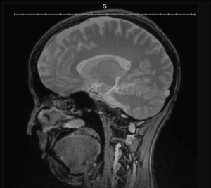
Double Inversion Recovery (DIR)
The Double Inversion Recovery sequence (DIR) has been advocated for detecting gray matter (GM) lesions due to its ability to enhance soft tissue contrast in the brain. It combines two inversion pulses in order to simultaneously suppress signals from tissues with different longitudinal relaxation times. In the brain, DIR allows to selectively image GM by nulling the signal from white WM and cerebrospinal fluid (CSF) at the time of the excitation pulse.
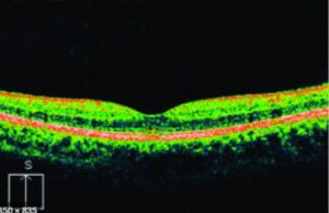
Ophthalmic Imaging
Within Multiple Sclerosis, it is possible to see structural changes in the retina, specifically within the Ganglion Cell Arteritis (GCA) and Nerve Fiber Layer (NFL). A combination of OCT, FP, and FAF may be appropriate for your trial to evaluate the retinal changes. The combination of Perceptive’s operational bench strength and global site/image processing capabilities with the expertise of Perceptive’s medical advisors make for an ideal study management structure.
Ophthalmology imaging endpoints have historically been serviced by academic core labs and though they offer the relevant expertise
in the modality and indication, they lack the infrastructure, validated quality systems and SOPs to manage a late phase study. Leveraging expertise across each lab, including Perceptive’s systems and export capability will reduce the added workarounds by the sponsor.
Case Studies
Advanced Imaging Analysis in Phase 2 Study
Perceptive Imaging played a vital role in a phase 2, double blind, randomized, multicenter trial in patients with relapsing-remitting multiple sclerosis including MTI analysis. During site standardization Perceptive supported the installation of the MTI specific software on the MRI scanners. 110 timepoint MTI datasets of 12 patients have been acquired, collected and processed. Structural MR image processing encompassed image pre-processing and linear spatial normalization, voxel classification. Lesion-based mean Magnetization Transfer Ratios (MTR) and voxel-based gray matter and white matter MTR histograms were derived for the MTR analysis.
Standard Imaging Analysis in Phase 3 Study
Perceptive Imaging played a vital role in a phase 3, double blind, randomized, multicenter trial in patients with relapsing-remitting multiple sclerosis. Based on the centrally assessed imaging data it was shown that the investigational drug significantly reduces MS lesion activity. The superiority of the investigational drug against interferon treatment was also demonstrated on clinical grounds (relapse rate reduction of52%). The trial was the largest phase 3 clinical trial program ever submitted to the FDA for a new MS drug at the time.
Experience
Perceptive Imaging’s experience is drawn from managing nearly 3,300 trials to date. Within this experience is our management of over 600 CNS protocols and over 30 MS studies of which 4 included advanced MRI.